Sunday, November 15, 2009

Fifth Observation of MicroAquarium


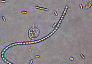
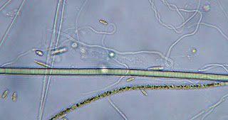
In this fifth and final observation of the MicroAquarium, Discomorpha, which is a flagellate and Anabeana, which is a diatom were identified (Pennak 1989). This observation was taken on November 12th. Decomposing green algae was observed, along with several other dead organisms, such as Phialina. Air bubbles and rapidly moving ciliates were still present in the aquarium. Amoebas, nematodes, and vortacella could still be seen, as well. No new pictures were taken with this week's observation. What organisms left in the aquarium were very small, and it was hard to get a well focused picture. The nematodes and the ciliates were the only organisms moving. The picture on the top is of Discomorpha, cyanobacteria, and diatoms, while the picture on the bottom contains the diatom, Anabeana. These pictures were taken during a previous observation.


Pennak RW. 1989. Freshwater Invertebrates of the United States Protozoa to Mollusca. New York (NY): John Wiley and Sons. p. 81.

Sunday, November 8, 2009

Fourth Observation of MicroAquarium

The MicroAquarium was observed for the fourth time on November 5th. Several clusters of dead organisms, such as Phialina were seen in the bottom of the aquarium. Phialina are torpedo shaped ciliates (Ward, Whipple 1918). The leaf residue had fully settled to the bottom of the aquarium and some of the water was gone. Air bubbles, ciliates, and halterians, which are flagellates were still present. A cyanobacteria with a heterocyst and decompsing green algae were also viewed. Some new organisms that were identified were Vorticella, Chania teres, and Coleochota irregularis (Forest 1954). All of these organisms were identified with the help of Dr. McFarland. More water was added at the end of my observation. From top to bottom the pictures are of Vorticella and Coleochaete irregularis.
Ward HB, Whipple GC. 1918. Freshwater Biology. NewYork (NY): John Wiley and Sons, Inc.
p. 272.
Forest HS. 1954. HandBook of Algae. Knoxville (TN): The University of Tennessee Press. p. 89.

Saturday, October 31, 2009

Third Observation of MicroAquarium


A week later, few changes can be seen in the MicroAquarium. Air bubbles, rapidly moving cialates, paranema, diatoms, and cyanobacteria are still present in the aquarium. One new organism, a Metazoa, which is a Nematode, was identified. Metazoa are long and thin organisms that make a whip like stroke in the water (Patterson 1992). Their appearance resembles that of a snake. Halteria and litonotus were both seen again in the aquarium. There did not appear to be any dead organisms. Some of the water in the aquarium had evaporated, so water was added. The pictures are of Paranema.


Patterson D.J. 1992. Free-Living Freshwater Protozoa A Color Guide. Washington (DC): Manson Publishing.

Saturday, October 24, 2009

Second Observation of MicroAquarium


The MicroAquarium was last observed on Thursday, October 22. Air bubbles were very noticeable in the aquarium, and the leaf residue from the water sample had fully settled to the bottom. The amount of organisms in the aquarium seemed to have increased. Approximately seven organisms were observed and identified in the Microaquarium. However, it is possible that more organisms are present. There were a lot of cialates moving very rapidly throughout the aquarium, and these ciliates were mainly halterians. Some other organisms that were seen were cyanobacteria, litonotus, diatoms, green alga, heterophyrs, and peranema (Patterson 1992). These organisms were identified with the help of Dr. McFarland. One pellet of "Atison's Beta Food" was added to the aquarium. "Atison's Beta Food" is made by Ocean Nutrition, Aqua Pet Americas. Their adress is 3528 West 500 South, Salt Lake City, Utah 84104. The ingredients in "Atison's Beta Food" are fish meal, wheat flower, soy meal, krill meal, minerals, vitamins, and preservatives. The breakdown of the ingredients in the food is 36% crude protein, 4.5% crude fat, 3.5% crude fiber, 8% moisture, and 15% ash. The picture on the top is a litonotus and the picture on the bottom is a halterian and a cyanobacteria.
Patterson DJ. 1992. Free-Living Freshwater Protozoa A Color Guide. Washington (DC): Manson Publishing.

Thursday, October 15, 2009

First Observation of Microaquarium

Water was obtained from bowl 2, along with some leaf residue by using a pipet. This water and leaf residue were exposed to partial shade and came from the French Broad River Water Shed at Seven Islands Wildlife Refuge in Knox County, Tennessee. The coordinates for this location were N35 56.742 W83 41.628 841 feet. Drops of water were put into the aquarium by using this pipet. There are many moving organisms in the aquarium, which seem to be swimming in the water. The organisms are single celled and are mainly non-green organisms, although there are two chlorophyll green plants in the aquarium, which were termed in the lab as plants A and B. Plant A is a moss with a scientific name of Amblystegium varium Lindb. The plant was found in a natural spring exposed to partial shade at Carter's Mill Park on Carter Mill Road in Knox County, Tennessee. The coordinates for this location were N36 01.168 W83 42.832. Plant B is a flowering plant and its scientific name is Utricularia vulgaris. This plant is carnivous. It was orginally retrieved from the south shore of Spain Lake on Camp Bella Air Road, just east of Sparta, Tennesee in White County. The coordinates for this location were N35o55 12.35" W088o20' 47.00. This plant was later grown in water tanks outside of the greenhouse at the Hesler Biology Building on the University of Tennessee campus. The plants were put into the aquarium by using forceps and a probe. The habitat preference of the organisms seems to be in the middle of the aquarium, and there are many of the same kind of organism. The leaf residue is mainly at the bottom of the aquarium. The date of this observation was October 13th.